High-speed multiphoton imaging of 420 fps with a resonant scanner |

The confocal microscope A1R, which has an excellent reputation for its high speed, superb sensitivity and high resolution, now offers multiphoton excitation imaging. The A1R MP incorporates these newly developed technologies in response to your requirement for the highest level of confocal and multiphoton imaging performance.
|
|
| |
Fast imaging with resonant scanner
|
| |
The resonant scanner is capable of imaging a wide area at a much higher speed than a non-resonant scanner, making it possible for 420-fps imaging, the world's fastest using point scanning technology. The NDD*1 for multiphoton microscopy makes it possible to image fast and deep through the thickest specimens. Nikon's optical pixel clock system allows more stable and more evenly illuminated imaging-even at high speeds.
|
|
*1 NDD (Non-Descanned Detector)
Unlike confocal imaging, where emitted light from the specimen passing through a pinhole is descanned before being detected, the A1R MP eliminates the need for a pinhole. By locating the NDD close to the back aperture of the objective, more of the scattered fluorescence emissions can be collected, improving the sensitivity of the instrument.
|
Visualization of intravital microcirculation
Blood cells in blood vessels within a living organism were excited by a femtosecond pulsed IR laser with the A1R MP's ultrahigh-speed resonant scanner, and their movements were simultaneously captured in three successive fluorescence images at 30 fps (30 msec), with three separate color channels.
Three fluorescent probes are simultaneously excited and imaged?nucleus (blue), endothelium (green), and plasma (red). The long-wavelength ultrafast laser in combination with the ultrahigh-speed resonant scanner effectively reduces photodamage and makes time resolved multiphoton imaging of biomolecules possible.
Image resolution: 512 x 512 pixels, Image acquisition speed: 30 fps, Objective: water immersion objective 60x
Imaged with the cooperation of: Dr. Satoshi Nishimura, Department of Cardiovascular Medicine, the University of Tokyo, TSBMI, the University of Tokyo, PRESTO, Japan Science and Technology Agency |
|
| |
Deep imaging with high-sensitivity Non-Descanned Detector |
| |
Nikon's newly developed 4-channel NDD* 1 for multiphoton microscopy allows imaging deep into a specimen.
A detector* 2 with very high sensitivity and a wider sensitive area than conventional models is placed close to the back aperture of the objective where image formation of the specimen takes place. This configuration improves the detection efficiency for scattered fluorescence, and enables the capture of signals from deep within a living organism more clearly and more reliably with higher S/N and less aberrations.
|
|
*2 Detecting as much scattered fluorescence as possible is important for observation of deep parts in a specimen. The detection depth varies depending on the detector sensitivity, size and installation position. |
|
| |
 |
|
 |
Figure 1
Image of deep areas of spinal cord primordia (neural tube) of a 13.5-day-old rat embryo
The entire embryo was cultured for approximately 24 hours after transfection with green fluorescent protein (GFP) by electroporation. A fixed cross-sectional slice of spinal cord was embedded in gel and two-photon excitation was conducted using pulsed IR laser. Observation of deep areas of over 600 μm was achieved.
Photographed with the cooperation of: Dr. Noriko Osumi, Dr. Masanori Takahashi, Division of Developmental Neuroscience, Tohoku University Graduate School of Medicine |
|
Figure 2
Image of fixed neuronal cells of mouse brain expressing eGFP captured using Non-Descanned Detectors (NDD)
Excitation with pulsed IR laser (920 nm) allowed higher S/N ratio imaging of deep brain tissue areas than with a confocal microscope. Optical sections of deep areas of over 600 μm in the Z-axis were achieved. A new CFI Apo LWD 40x WI λS objective was used.
Photographed with the cooperation of: Dr. Satoru Kondo, Department of Cellular Neurobiology, Graduate School of Medicine, the University of Tokyo |
| |
Sharp, bright imaging with high NA objectives
|
| |
The new water immersion objectives offer superior chromatic aberration correction throughout a wide wavelength range and high transmission covering from near-ultraviolet to near-infrared range, made possible by Nikon's proprietary Nano Crystal Coating* 3 .
CFI Plan Apo IR 60x WI with an NA of 1.27?the world's highest for a 60x water immersion objective?offers high-contrast imaging with excellent resolution and brightness.
*3 An ultra-low refractive index film that was initially developed for Nikon's IC steppers. With its coarse structure?nano particles are arranged in a spongy composition?thus the Nano Crystal Coating boosts the transmission of light over a wide spectral wavelength range.
|
|
|
Objectives for multiphoton microscopy
| CFI Apo LWD 40x WI λS |
NA 1.15, W.D. 0.59-0.61 mm  |
| CFI Apo 40x WI λS |
NA 1.25, W.D. 0.20-0.16 mm |
| CFI Plan Apo IR 60x WI |
NA 1.27, W.D. 0.18-0.16 mm |
| CFI Plan Fluor 20x A MI |
NA 0.75, W.D. 0.35 mm |
|
| Nano Crystal Coat-deposited |
|
 |
Fixed spinal cord primordia (neural tube) of a GFP transfected 13.5-day-old rat embryo after a 24-hour whole embryo culture
Neuroepithelial cells and commissural axon of migrating interneurons can be clearly observed.
Photographed with the cooperation of: Dr. Noriko Osumi, Dr. Masanori Takahashi, Division of Developmental Neuroscience, Tohoku University Graduate School of Medicine
|
 |
Fixed neuronal cells of mouse brain expressing eGFP
Excitation with pulsed IR laser (920 nm) allowed higher S/N ratio imaging of deep brain tissue areas than with a confocal microscope.
Photographed with the cooperation of: Dr. Satoru Kondo, Department of Cellular Neurobiology, Graduate School of Medicine, the University of Tokyo |
| |
Precise, high-speed, high-contrast unmixing*4 |
| |
In addition to spectral detection and unmixing by the 32-channel detector, unmixing by the 4-channel NDD* 1 for multiphoton microscopy is also realized. Images captured with a resonant scanner can be unmixed, so clear and high-contrast images of areas deep within a thick specimen can be captured in ultrahigh speed.
|
|
*4 Fluorescence from a specimen stained with multiple fluorescence probes is separately detected by wavelength, and the brightness and the spectral response of the fluorescence probes can be measured. This method allows easy unmixing of fluorescence probe combinations?such as GFP and YFP?that cannot be unmixed by conventional methods.

Image of specimen stained with Alexa 488, MitoTracker Red and DAPI was captured with spectral detector during two-photon excitation and unmixed. Excitation with infrared wavelength reduces damage to live cells. |
| |
 |
Multiphoton laser beam alignment with a single click
When the multiphoton laser wavelength or group velocity dispersion precompensation is changed, the multiphoton laser beam positional pointing may drift, resulting in uneven intensity of images, or a slight misalignment of fluorescence images that are produced by a visible light laser and a multiphoton laser.
Since the multiphoton laser is invisible to the eye, the laser beam alignment has been difficult and potentially dangerous for users, especially at wavelengths longer than 800 nm. Nikon A1R MP's newly developed auto laser alignment function automatically corrects multiphoton laser misalignments with a single click. |
|
 |
| |
|
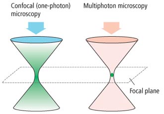
Principle of multiphoton excitation
The multiphoton confocal microscope enables excitation by simultaneous absorption of two or more near infrared photons by a single fluorescent molecule. When two photons are absorbed simultaneously into a single fluorescent molecule (two-photon excitation), the excitation efficiency is proportional to the square of the excitation light intensity.
In order to achieve multiphoton excitation, a pulsed beam with high photon density or flux of photons is used. Because the laser beam is delivered in very short (femtosecond) pulses and is converged on the focal point through an objective lens, the probability of simultaneous absorption of two photons becomes high enough to be useful for imaging.
The intensity of the laser beam converged by the objective lens decreases in inverse proportion to the square of the distance from the focal plane. Therefore, the efficiency of two-photon excitation decreases inversely with the fourth power of the distance from the focal plane. As a result, only a fluorescenc///e molecule located within the diffraction-limited focal volume of the objective lens is excited and emits fluorescence.
Because of the small amount of absorption and scattering by a specimen, the near infrared light used for excitation penetrates deep into thick tissue without losing much of its strength. Also, the excitation range is only within the diffraction-limited focal volume of the objective lens. Therefore, photodamage to a specimen can be minimized, making it suitable for observation of whole organisms in vivo, live cells and live tissues. The combination of the group velocity dispersion precompensation system incorporated in the multiphoton laser and the multiphoton detector (NDD) allows fluorescence imaging of deeper areas of a specimen than standard confocal technique.
|
|
Transition of energy levels of fluorescence Excitation area in confocal microscopy and multiphoton microscopy
molecule
|
| |
|